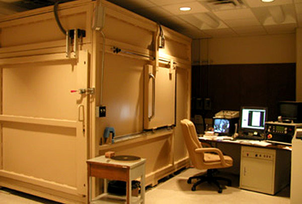
CT scanning equipment. At left is the lead box that houses the scanner used for the Ceremonial Cave specimens. Image courtesy of Jackson School of Geosciences, University of Texas. |
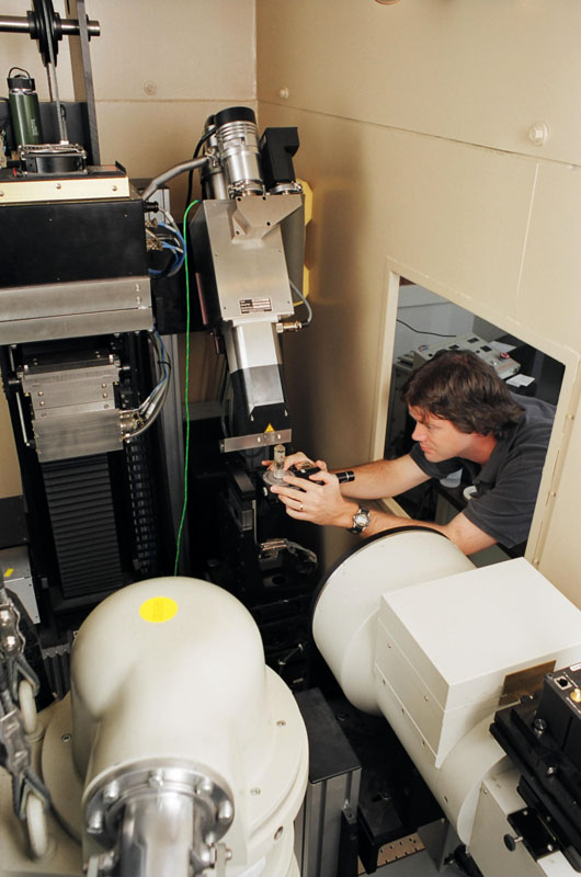
Rich Ketcham, laboratory director, prepares a specimen for scanning. The system on right is the FeinFocus, and the X-ray source is byond, with green wire. The detector is in the forefront. Image courtesy of Jackson School of Geosciences, University of Texas. |
High resolution, non-destructive, X-ray computed tomography density maps were made at the High-Resolution X-ray Computed Tomography Facility in the Department of Geological Sciences in the Jackson School of Geosciences, University of Texas at Austin. In essence, these scans enable us to see "underneath" the hafting materials and to view the hidden stem portions of these hafted chipped-stone dart points. This is particularly important in dart point typology because the stem, or basal portion, typically is the more diagnostic and temporally sensitive. Unlike the blade portions of dart points, which may have been altered by use-damage and re-sharpening, the stem is most likely to have remained in its original form, protected by the haft covering. Dart point styles and shapes are useful in tracing cultural patterns over time.
Scanning of the hafted dart points in this study involved taking approximately 500 CT slices, from the tips of the points to slightly beyond the base of the points (where only the wood foreshaft was present). From this raw digital imagery, rotating "movies" with 3-dimensional details were generated for each chipped-stone point. Specimens were scanned by Matthew Colbert and images processed by Jessie Maisano.
These can be viewed as Quicktime movies and as selected cross-section slices in the Specimen pages of this exhibit. The 3-D images of the chipped-stone dart point and the haft portion of the dart shaft have been colorized to represent the weapon in its original state.
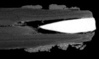
CT scan showing lateral view of dart point seated in haft. |
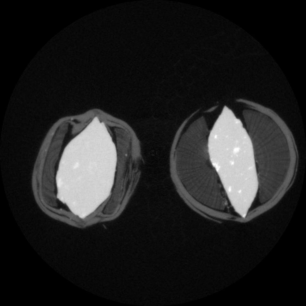
Cross-section slices of dart point, shown at different locations within the haft. Images such as these reveal details of foreshaft construction and types of materials used, such as flower stalk, wood or cane. |
